|Articles|May 5, 2020
Can Artificial Intelligence Predict Glaucoma Progression?
Author(s)Gianna Melillo
A new test, supported by an artificial intelligence (AI) algorithm, can detect glaucoma progression a year and a half earlier than widely used optical coherence tomography retinal imaging technology, according to a study published in Expert Review of Molecular Diagnostics.
Advertisement
A new test, supported by an artificial intelligence (AI) algorithm, can detect
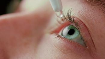
The Detection of Apoptosing Retinal Cells (DARC) test involves injecting fluorescent dye into the bloodstream, which then attaches to retinal cells. The dye illuminates cells in the process of apoptosis, enabling visual detection during eye examinations. Higher numbers of damaged cells indicate a higher DARC count.
“Glaucoma is a progressive and slowly evolving ocular neurodegenerative disease that it is the leading cause of global irreversible blindness, affecting over 60.5 million people. It is predicted to double by 2040 as the aging population increases,” the researchers said. “A key objective in glaucoma is to identify those at risk of rapid progression and blindness.”
When diagnosing eye diseases, some specialists may disagree on what exactly is presented in eye scans. To address potential discrepancies, the researchers created an AI algorithm using convolutional neural networks (CNNs) to effectively and objectively classify glaucoma.
A phase 2 clinical trial testing DARC and the algorithm included 20 individuals with progressing glaucoma and 40 healthy control subjects. The researchers collected baseline infrared auto-fluorescent images prior to administering fluorescent liquid. Following a single dose of 0.4 mg of ANX776 injection, images were taken after 15, 120, and 240 minutes. The investigators recorded average images from sequences of 100 frames at each time point. These images were then fed into the algorithm.
“For the CNN-training, 73 control eyes at baseline and 120 minutes were available for the analysis. Similarly, of the 20 glaucoma patients who received intravenous ANX776, images were available for 27 eyes at [the] baseline and 120 time-points,” authors said.
A total of 58,730 spot candidates (apoptotic cells) were taken from baseline images, and 70% of these spots were used to train the algorithm. The remaining 30% were used to validate classifications. In addition, retinal images of the remaining 50% of control patients were used to test the classification accuracy (48,610 candidate spots), according to the researchers.
Next, the researchers fed the algorithm images from patients with glaucoma, captured both at baseline and at 120 minutes. Manual observers also compared spots for each image.
“The criteria for spots used in the automated application to train and compare the systems was when there was concordance of 2 or more observers," they noted.
After 18 months, the researchers followed up with individuals to see whether their eye health had deteriorated and whether the algorithm effectively predicted glaucoma progression. Using the CNN-enabled algorithm, thye found “DARC count was…significantly higher in patients who were later found to be progressing at 18 months (mean, 26.13; 95% CI, 9.41-42.84) compared to those who were stable (mean, 9.71; 95% CI, 5.68-13.75; P = .0044).”
Compared with manual observation accuracy, the CNN-aided AI was superior in predicting glaucoma progression. In addition, the researchers discovered a reliable biomarker indicating risk of progressive glaucoma, in that after 18 months, no stable eyes had a CNN DARC count above 30.
Data revealed “maximal sensitivity (85.7%) and specificity (91.7%) were achieved above a DARC count of 24, with an area under the curve (AUC) of 0.88 and likelihood ratio of 8.57 with the CNN algorithm as opposed to the manual observer with maximal sensitivity (71.4%) and specificity (87.5%) above a DARC count of 11, an AUC of 0.79, and likelihood ratios of 4.76.” In comparison, OCT “has been found to have a sensitivity and specificity of 83% and 88%, respectively, for detecting significant [retinal nerve fiber layer] abnormalities in addition to good repeatability.”
The investigators hope the technology can aid in accelerating clinical trials for glaucoma treatments and that it may eventually be used in diagnostics.
"We have developed a quick, automated, and highly sensitive way to identify which people with glaucoma are at risk of rapid progression to blindness,”
In response to the promising results, the AI-supported technology has been approved by the United Kingdom’s Medicines and Healthcare products Regulatory Agency, along with the United States’ FDA as an “exploratory endpoint for testing a new glaucoma drug in a clinical trial.”
Future longitudinal studies are needed to further validate the findings. Currently, researchers are applying the test to rapidly detect cell damage caused by conditions like age-related macular degeneration, multiple sclerosis, and dementia.
DARC is also being tested in individuals with lung disease, and investigators hope it may aid in assessing individuals with breathing difficulties resulting from coronavirus disease 2019 (COVID-19).
Reference
Normando EM, Yap TE, Maddison J, et al. A CNN-aided method to predict glaucoma progression using DARC (Detection of apoptosing retinal cells) [published online May 3, 2020]. Expert Rev Mol Diagn. doi: 10.1080/14737159.2020.1758067.
Advertisement
Related Content
Advertisement


Advertisement
Advertisement
Trending on AJMC
1
FDA Approves Enlicitide, First Oral PCSK9 for High Cholesterol
2
SYNCHRONIZE-1 Through a Hepatologist's Lens: Liver Fat Reduction, Visceral Fat, and What Comes Next
3
Beyond GLP-1: The Biological Case for Dual Incretin Therapy in MASH
4
Stem Cell Transplant Outperforms DMTs in Drug-Resistant Relapsing MS
5