|Articles|September 9, 2022
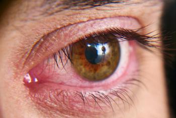
Fluorescein Angiography Predominantly Peripheral Lesions Associated With Worsening of Diabetic Retinopathy
Author(s)Julia Bonavitacola
A prospective, multicenter, longitudinal observational study found that diabetic retinopathy worsening could be associated with fluorescein angiography predominantly peripheral lesions over 4 years.
Advertisement
Ultra-widefield (UWF) images can identify predominantly peripheral lesions (PPLs), which may in turn be used to identify eyes that are at risk of
The study was conducted at 37 clinical sites in the United States and Canada. All participants were 18 years and older, had type 1 or type 2 diabetes, and had at least 1 eye with nonproliferative DR (NPDR) that was confirmed using the Early Treatment Diabetic Retinopathy Study’s (ETDRS) 7-field grading. There were 388 participants enrolled between February 2015 and December 2015, and all data were analyzed from May 2020 to June 2022. There were 544 study eyes from 367 participants. Theedian (IQR) age of the participants was 62 (53-69) years, 50% were women, and 68% were non-Hispanic White.
Follow-up visits were annual for the 4 years of the study. Images were acquired at each visit, and the start of treatment for DR was at the investigator’s discretion. Disease progression was defined as a worsening by 2 or more steps on the Diabetic Retinopathy Severity Scale assessed in the ETDRS fields, from the UWF-color images or receipt of DR treatment.
There were 221 eyes (41%) that had PPLs present on UWF-color and 247 (46%) that had PPLs on UWF-FA. There were 136 eyes (25%) that had PPLs present in both images, 111 (20%) that had FA PPLs only, 85 (16%) that had color PPLs only, and 210 (39%) that did not have PPLs. Hemorrhages and/or microaneurysms were the most common types of PPLs, having been detected in 180 of 221 eyes (81%) on UWF-color and 225 of 247 eyes (91%) on UWF-FA.
Treatment for DR or diabetic macular edema (DME) was started in 18% of all eyes, with 11% of this group receiving for DR and 14% for DME. The total proportion of eyes that worsened over 4 years was 40% (95% CI, 36%-45%) overall, 45% (95% CI, 37%-54%) in eyes with baseline mild NPDR, 40% (95% CI, 32%-49%) with moderate NPDR, 26% (95% CI, 17%-38%) with moderately severe NPDR, and 43% (95% CI, 34%-53%) with severe or very severe NPDR in UWF-color images.
When the results were stratified by baseline PPL status, 38% (95% CI, 31%-45%) of eyes with color PPLs and 43% (95% CI, 37%-49%) without color PPLs had disease worsening (HR, 0.78; 95% CI, 0.57-1.08). The primary outcome rate was 50% (95% CI, 44%-57%) in eyes with FA PPLs and 31% (95% CI, 25%-38%) in eyes without FA PPLs.
Seventeen percent of eyes (95% CI, 12%-23%) with color PPLs and 26% (95% CI, 21%-32%) of eyes without color PPLs examined in this study developed proliferative DR or received DR treatment (HR, 0.90; 95% CI, 0.57-1.44). The percentage was higher in eyes with FA PPLs than those without (24% vs 20%; HR, 1.60; 95% CI, 1.05-2.45).
There were some limitations to this study. The last year of follow-up occurred during the COVID-19 pandemic and was therefore affected by it, mostly through the inability to comply with study visits. There was also only a 77% completion rate in the 4 years, excluding deaths, and newer UWF imaging technology has been developed to detect color PPLs since the start of this study.
The researchers concluded that the presence of FA PPLs was associated with a significantly greater risk of worsening in the disease or treatment over 4 years.
“These results suggest use of UWF-FA to evaluate retina peripheral to standard ETDRS fields improves the ability to predict disease worsening in NPDR eyes. These findings support use of UWF-FA for future DR staging systems and clinical care to more accurately determine prognosis in NPDR eyes,” the authors wrote.
Reference
Marcus DM, Silva PS, Liu D, et al. Association of predominantly peripheral lesions on ultra-widefield imaging and the risk of diabetic retinopathy worsening over time. JAMA Ophthalmol. Published online August 18, 2022. doi:10.1001/jamaophthalmol.2022.3131
Advertisement
Related Content
Advertisement


Advertisement
Advertisement
Trending on AJMC
1
AI Chatbots Score Below 28% on Colorectal Cancer Questions
2
5 Notable FDA Approvals From the First Half of 2026
3
FDA Approves Isatuximab On-Body Injector, Raising Anti-CD38 Competition in Myeloma
4
Michigan Cyclospora Outbreak Surpasses 1500 Cases as Investigation Continues
5