News
Article
Key MRI Features That Likely Correlate With STS Malignancy Grades
Author(s):
Investigators from Germany conducted a systematic review of different MRI features in soft tissue sarcoma (STS) that are used to infer tumor histological grade.
Six characteristics used to assess tumor histological grade in soft tissue sarcoma (STS) were shown to have significant associations with STS malignancy grade in a systematic review and meta-analysis of 14 studies conducted by 2 investigators from German, according to new data published in European Journal of Radiology.1
Those 6 characteristics are tumor size, tumor margin, necrosis, peritumoral edema, contrast enhancement, and polycyclic/multilobulated tumor configuration, and the studies that evaluated them encompassed 705 patients.
“The gold standard for determining the grade of malignancy is core needle biopsy. However, biopsy may lead to underestimation of the malignancy grade due to intratumoral heterogeneity,” the study authors wrote. “Simplifying the reporting of STS on MRI would be advantageous, especially regarding estimating the malignancy grade, consecutively affecting patients’ prognosis.” However, they also noted the lack of systematic reviews in the space on using MRI to grade STS, which is why they conducted their systematic review.
PubMed and MEDLINE were searched for published investigations of correlations between STS MRI findings and tumor grade. They used the keywords MRI, magnetic resonance imaging, sarcoma, grade, grading, and FNCLCC (Federation Nationale des Centres de Lutte Contre le Cancer). FNCLCC, the study authors noted, is a common grading system for STS malignancy; it uses tumor differentiation, mitotic count, and tumor necrosis for histological grading.2


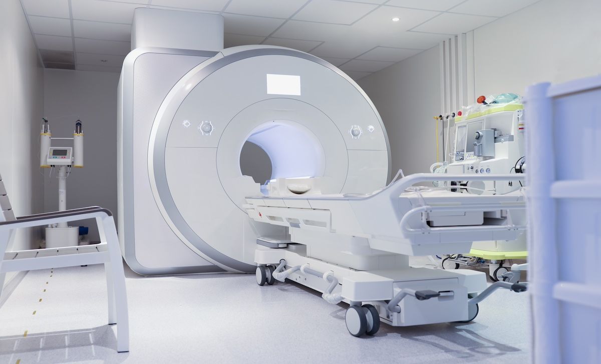
The authors of this study note that although core needle biopsy is the gold standard for determining tumor malignancy grade, the potential for intratumoral heterogeneity with the method is great | Image Credit: megaflopp-stock.adobe.com
In addition to the characteristics already mentioned, heterogeneity in T2w images, signal intensity in T1w images/hemorrhage, peritumoral contrast enhancement, tumoral diffusion restriction, signal intensity in T2w images/necrosis, internal low signal intensity septations, deep tissue layer, periosteal reaction, cortex erosion, marrow extension, joint extension, neurovascular encasement, vascular occlusion, contrast enhancement, apparent diffusion coefficient (ADC) value, tail sign, and bone invasion were evaluated as MRI features able to confer STS tumor histological grade. Only observational studies were included in this analysis, and there ultimately were 14 full-text articles included.
Overall, the investigators found a high degree of heterogeneity in these studies, with the numbers of MRI features evaluated ranging from 1 to many, while patient populations ranged from 12 to 130. Correlation significance was demonstrated by overall P values and total studies that determined that characteristic to have a significant correlation at being able to infer STS histological grade.
The following results were seen for each significant factor:
- Tumor size was significant in 4 of 9 studies, and ranged from P = .015 (n= 51) to P = .81 (n = 36)
- Tumor margin was significant in 3 of 5 studies, and ranged from P < .001 (n = 95) to P = .93 (n = 36)
- Necrosis was significant in 3 of 8 studies, and ranged from P = .004 (n = 50) to P = 0.65 (n = 95)
- Peritumoral edema was significant in 3 of 6 studies, and ranged from P = 0.002 (n = 130) to P = 0.337 (n = 40)
- Contrast enhancement was significant in 3 of 3 studies, and ranged from P < .01 (n = 50) to P = .019 (n = 51)
- Tumor configuration/shape was significant in 1 study (P = .008; n = 71)
Less significance was attributed to these characteristics:
- Heterogeneity in T2w images (P = .003 [n = 130] to P = .202 [n = 40])
- Signal intensity in T1w images (P = .02 [n = 130] to P = .5 [n = 31])
- Peritumoral contrast enhancement (P < .001 [n = 95] to P = .253 [n = 51])
- ADC mean (P = .01 [n = 51] to P = .28 (n = 36])
“Our review revealed that there is no common consensus about what MRI features assist in inferring the STS grade,” the authors wrote. “Due to the heterogeneity and rarity of STS, simplification and further insight into imaging characteristics are necessary. The grading of STS is crucial for therapy planning.”
Further research is still needed to improve consensus on the relevance of these features, they concluded.
References
1. Schmitz F, Sedaghat S. Inferring malignancy grade of soft tissue sarcomas from magnetic resonance imaging features: a systematic review. Eur J Radiol. 2024:177:111548. doi:10.1016/j.ejrad.2024.111548
2. National Cancer Institute. Childhoos soft tissue sarcoma treatment: FNCLCC histologic grading system. PDQ Cancer Information Summaries. Accessed June 13, 2024. https://www.ncbi.nlm.nih.gov/books/NBK65923.1/table/CDR0000062934__749/?report=objectonly


A Novel Approach to Chronic GVHD With Axatilimab: Dr Daniel Wolff