|Articles|March 17, 2020
MRIs Find Abnormalities in Central Vestibular Cortex of Some Patients With Migraine
Author(s)Gianna Melillo
Magnetic resonance imaging (MRI) scans of patients with vestibular migraine reveal abnormalities in the central vestibular cortex, according to a study published in Brain and Behavior.
Advertisement
Patients with vestibular migraine (VM) exhibit abnormalities in the central vestibular cortex, according to a
In addition, the study found correlations between dizziness severity and gray matter volume (GMV) in core regions of the vestibular cortex. Combined, these findings suggest a pathophysiological role of the core vestibular regions in patients with VM.
VM is characterized as “vestibular symptoms, such as recurrent episodes of vertigo, and migrainous symptoms including headache, photophobia, and phonophobia,” according to researchers. Women tend to suffer from VM 2 to 3 times more frequently than men.
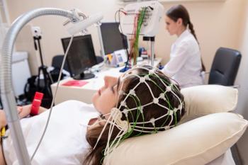
Researchers conducted magnetic resonance imaging of 20 patients with VM and 20 healthy controls to estimate GMV and assess the relationship between clinical parameters and morphometric abnormalities in patients with VM. The scans were performed between days 3 to 7 after a VM attack, and all patients were free of migraines and vertigo on the day of the scan.
When compared with controls, patients with VM exhibited decreased GMV in the following regions:
- Prefrontal cortex
- Posterior insula-operculum regions
- Inferior parietal gyrus
- Supramarginal gyrus
Study participants also completed questionnaires including the Dizziness Handicap Inventory (DHI) and the Migraine Disability Assessment. A correlation analysis found DHI scores were negatively correlated with the volume of the left posterior insula-operculum regions (P = .04).
Researchers highlighted the reduction of GMV in the posterior insula-operculum in patients with VM, and reduced GMV in the operculum, which is adjacent to the posterior insula. “The posterior insular—opercular regions are believed to contribute to pain transmission as the receiving areas of the spinothalamic system, which remains the crucial part of the pain network…Therefore, our findings of GMV loss in these regions might reflect the important role of these transmission circuitry impairments in the pathophysiology of VM,” they said.
Due to the small sample size, lack of distinguishing between migraine with aura and migraine without aura, and other limitations, the study authors noted that future studies will aid in further understanding the pathophysiology of VM. They suggested future studies combine structural and functional analyses to do so.
Reference
Zhe X, Chen L, Zhang D, et al. Altered structure of the vestibular cortex in patients with vestibular migraine [published online March 10, 2020]. Brain Behav. doi: 10.1002/brb3.1572.
Advertisement
Related Content
Advertisement


Advertisement
Advertisement
Trending on AJMC
1
FDA Approves Enlicitide, First Oral PCSK9 for High Cholesterol
2
SYNCHRONIZE-1 Through a Hepatologist's Lens: Liver Fat Reduction, Visceral Fat, and What Comes Next
3
Beyond GLP-1: The Biological Case for Dual Incretin Therapy in MASH
4
Stem Cell Transplant Outperforms DMTs in Drug-Resistant Relapsing MS
5