|Articles|July 27, 2021
Negative Pressure Wound Therapy Reduced Amputation Risk for Patient With Diabetic Wound
Author(s)Skylar Jeremias
A patient with a diabetic foot wound who underwent negative pressure wound therapy experienced good healing, reduced amputation risk, and no wound infection, suggesting the treatment method could be effective in other patients with similar wounds.
Advertisement
A patient who received negative pressure
The report lends further support to previously published research that has shown NPWT to be effective at decreasing healing time, reducing ulcer area, and increasing healing rates of ulcers. NPWT has also been shown to aid patients with foot ulcers in achieving complete ulcer closure better than advanced moist wound therapy.
NPWT, a wound treatment used for open wounds since the 1940s, involves a vacuum-sealed drain and vacuum-sealed closure to create a localized controlled negative pressure environment for a wound. Research has demonstrated that NPWT is a promising treatment method for complex diabetic foot wounds, especially for chronic wounds, and may be effective when used as an adjuvant treatment for temporary closure and preparation for wound beds prior to surgery.
The investigators analyzed the results of applying NPWT to a 40-year-old male patient with type 2 diabetes who presented at the Department of Surgery and Anatomy at University of São Paulo in Ribeirão Preto, Brazil, with plantar perforating disease on his right side, which was not treated by a specialist. The patient was obese, sedentary, and dyslipidemic upon hospital presentation.
The wound worsened with hyperemia in the instep and produced discharge of purulent secretion from the side of the foot. The patient also experienced fever, chills, and pain in the lower limb. After 7 days without a satisfactory wound response, the patient experienced increased necrosis on the leg, a foul odor, pain, edema, and purulent secretion discharge and was referred to the hospital.
Upon admission, the patient was administered intravenous antibiotic therapy once daily. Doctors performed vascular surgery to assess the extent of the wound and vascularization of the right lower limb, determining that palpation of the distal pulses was not possible.
The patient underwent debridement of the right lower limb, and a large amount of necrotic tissue and purulent secretion was removed. The injury was severe enough that several tendons and muscles in the dorsum of the foot and leg were resected, leaving bone exposure.
The wound was thoroughly cleaned using saline and physicians implanted a Renesys Smith Nephew dressing with negative pressure at 120 mm Hg. The first dressing change occurred after 3 days, and the wound showed significant improvement. However, a large amount of discharge remained in the anterior compartment of the leg.
With each vacuum dressing change and saline cleaning, which occurred every 3.5 days on average based on the amount of secretion drainage, the investigators noticed increases in granulation tissue and reduction in bone exposure.
Over the course of 70 days, 20 dressing changes were performed, and continuous negative pressure of 120 mm Hg was maintained. After complete coverage of granulation tissue was achieved without infection, hospital staff performed elastic suturing in the leg to reduce the size of the dermis and epidermis graft, which was not possible in the dorsum of the foot. Vacuum dressing was reinstalled after the wound was cleaned.
After 71 total days in the hospital, the patient underwent a dermal matrix implant procedure, during which an epidermis graft was removed from the thigh and placed on the wound. The patient experienced good recovery and no signs of necrosis around the graft, and was discharged from the hospital 22 days after the grafting procedure.
Five months after the grafting, the areas that experienced tissue loss had recovered well and gradually reduced in size. The patient’s foot still had sensation and motor function. Also, the patient’s diabetes was controlled after entering a food reeducation program, and they underwent motor physiotherapy to maintain foot function.
Reference
Fabiano de Oliveira Leite T, Ribeiro da Silva E, Joviliano EE. Effect of negative pressure wound therapy for legs in complex wound diabetic patients: therapeutic challenge and review. SAGE Open Med Case Rep. 2021;9:1-6. doi:10.1177/2050313X2211025920
Advertisement

Advertisement
Advertisement
Trending on AJMC
1
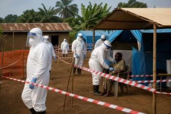
Ebola Cases Top 1700 as Unpaid Health Workers Strike
2
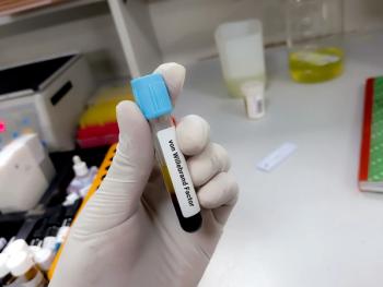
FDA Expands Approval for VWD Prophylaxis in Children Younger Than 6
3
Upper Payment Limit Implementation Risks for Patients, Pharmacies, and Providers
4
Gene Therapy Could Ease Wet AMD's Injection Burden: Carl Regillo, MD
5