|Articles|September 24, 2020
Tool Holds Potential for Biomarker-Based Diagnoses of Adolescent Concussion
Author(s)Gianna Melillo
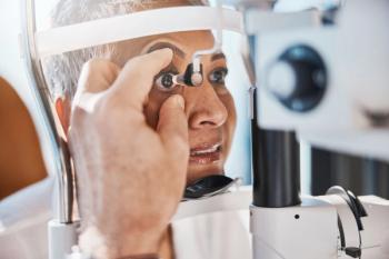
For the first time, researchers demonstrated that quantitative pupillary light reflex (PLR) metrics can be used to differentiate concussed adolescent athletes from healthy athletes.
Advertisement
For the first time, researchers demonstrated that quantitative
“Convergence and accommodation deficits after concussion predict prolonged recovery, while exercise intolerance is another manifestation of autonomic dysfunction after sports-related concussion (SRC),” authors explained. “The PLR is involved in both convergence and accommodation, driven by the parasympathetic system for constriction and the sympathetic system for dilation.”
Concussion diagnosis remains clinical and is based on a history of injury and onset of a subjective and nonspecific constellation of symptoms. However, whereas adults demonstrate decreasing pupil size with increasing age, children, specifically boys aged 12 to 18 years, have larger maximum pupil diameters, slower maximum constriction velocities, and smaller percentage constriction compared with younger children aged 6 to 11 years. These characteristics hinder objective diagnoses of concussion in this age group.
In this prospective observational cohort study, researchers aimed to determine whether differences in quantitative PLR metrics could serve as an objective physiologic biomarker for adolescent SRC.
Participants aged 12 to 18 years were enrolled in the study between August 2017 and December 2018. A total of 98 athletes with a diagnosis of SRC were recruited from a concussion program and a private suburban high school. One hundred and thirty-four healthy controls were also recruited from the high school.
Each participant with concussion underwent a PLR assessment within 28 days of injury with an assessment completed at a median (interquartile range) of 12 (5-18) days following injury.
“Exclusion criteria for both cases and controls included a concussion within 1 month of injury or preinjury assessment, ongoing chronic post-concussion symptoms, eye trauma, any ocular or neurologic condition, or medication that could affect pupillary responses,” researchers wrote.
A Neuroptics PLR-300 handheld, infrared, automated monocular pupillometer was used to measure pupillary dynamics including maximum pupil diameter, minimum pupil diameter, percentage pupil constriction, latency, peak and average constriction velocity, average dilation velocity, peak dilation velocity, and T75 (time for pupil redilation from minimum diameter to 75% maximum diameter). The device captured dynamic responses 32 times per second analyzing a continuous 5-second digital video of the pupillary response to light.
Among athletes with concussion, 8 of 9 PLR metrics were significantly greater compared with controls:
- Maximum pupil diameter: 4.83 mm vs 4.01 mm; difference, 0.82 (99.44% CI, 0.53-1.11)
- Minimum pupil diameter: 2.96 mm vs 2.63 mm; difference, 0.33 (99.4% CI, 0.18-0.48)
- Percentage constriction: 38.23% vs 33.66%; difference, 4.57 (99.4% CI, 2.60-6.55)
- Average constriction velocity: 3.08 mm/s vs 2.50 mm/s; difference, 0.58 (99.4% CI, 0.36-0.81)
- Peak constriction velocity: 4.88 mm/s vs 3.91 mm/s; difference, 0.97 (99.4% CI, 0.63-1.31)
- Average dilation velocity, 1.32 mm/s vs 1.22 mm/s; difference, 0.10 (99.4% CI, 0.00-0.20)
- Peak dilation velocity: 1.83 mm/s vs 1.64 mm/s; difference, 0.19 (99.4% CI, 0.07-0.32)
- T75: 1.81 seconds vs 1.51 seconds; difference, 0.30; 0.10-0.51
Researchers also found that girls with concussion exhibited longed T75 (1.96 seconds vs 1.63 seconds; difference, 0.33 [95% CI, 0.02-0.65]) compared with their male counterparts. Healthy control individuals exhibited diminished PLR metrics after exercise, such as a smaller maximum pupil size, indicating a potential fatigue effect.
A total of 6% of control participants and almost 11% of athletes with concussion did not have adequate data collected due to either artifacts or inability to complete the assessment secondary to symptoms. Participants were also predominately white, limiting generalizability of results. Lower luminosity or more specific instructions prior to assessment may improve these rates in future studies.
Investigators hope the findings lead to a future assessment tool that can be used both in sports and urgent care settings. “The association of concussion with PLR metrics appears robust, with significant differences between athletes with concussion and control participants in all metrics except for latency,” researchers concluded.
Reference
Master CL, Podolak OE, Ciuffreda KJ, et al. Utility of pupillary light reflex metrics as a physiologic biomarker for adolescent sport-relted concussion. JAMA Ophthalmol. Published online September 24, 2020. doi:10.1001/jamaophthalmol.2020.3466
Newsletter
Stay ahead of policy, cost, and value—subscribe to AJMC for expert insights at the intersection of clinical care and health economics.
Advertisement
Related Content
Advertisement


Advertisement
Advertisement
Trending on AJMC
1
TrumpRx Launch Brings Savings—and Uncertainty
2
How Do GLP-1 Agonists Fit Into Stroke Care?
3
Addressing Disparities at Every Step of the Stroke Care Pathway: Luke Messac, MD, PhD
4
5 Things to Know About the Oral GLP-1 Era
5


