|Articles|March 24, 2020
OCTA Device Successfully Used on Extreme Low Birth Weight Neonates
Author(s)Gianna Melillo
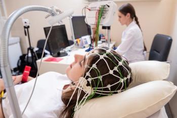
Researchers were able to effectively use an arm-mounted optical coherence tomography angiography (OCTA) device to learn information about the developing fovea in extreme low birth weight (ELBW) neonates, according to a study published in the American Journal of Ophthalmology.
Advertisement
Researchers were able to effectively use an arm-mounted optical coherence tomography angiography (OCTA) device to learn information about the developing fovea in extreme low birth weight (ELBW) neonates, according to a
Infants born weighing less than 1000g or before 27 weeks of gestational age (GA) were considered ELBW neonates. Effective use of an OCTA device to measure the foveal avascular zone (FAZ) area and retinal thickness introduces the possibility for researchers to “study the effects of ELBW, peripheral retinopathy of prematurity (ROP) and ROP treatment on foveal development.”
ELBW infants have higher risks of developing ROP and by extension, childhood blindness. This observational case series, performed at the Ronald Reagan Medical Center, is the first of its kind to use an arm-mounted OCTA device in neonates to study FAZ area associated with prematurity.
Researchers obtained images of 13 eyes from 7 infants with an average GA at birth of 25 weeks. All infants underwent ROP treatment ROP treatment in at least 1 eye. In addition, average birthweight (BW) was 615 g. Researchers drew the following conclusions from the images:
- There was no correlation between FAZ area and GA (superficial capillary plexus [SCP] P = .44, deep capillary plexus [DCP] P = .66)
- There was no correlation between FAZ area and BW (SCP P = .73, DCP P = .83)
- There was a positive correlation between FAZ area in both the SCP and DCP and the ratio of the outer to inner retinal layer thickness (P = .02; P = .02)
- The FAZ area in the SCP corresponded to a thinner inner retina (P = .008)
- A thicker outer retina corresponded with older PMA (P = .02)
- A thicker inner retina corresponded to a higher GA (P = .01) and higher PMA (P = .03)
“Our study highlights that there are differences in the development of the FAZ in infants born preterm versus those born at term,” researchers said. They also note the study indicates an OCTA device may be “very useful to investigate foveal development in ELBW infants.”
Futures studies should include a larger sample size and pre-and post- treatment images of infants’ eyes. Additional studies could also “address the question of if and how laser and anti- vascular endothelial growth factor treatment for peripheral ROP affects foveal maturation by imaging before and after treatment,” researchers said.
Reference
Kothari N, Chu A, Huang JM. Arm-mounted optical coherence tomography angiography in extremely low birth weight neonates with retinopathy of prematurity [published online February 24, 2020]. Am J Ophthalmol. doi: 10.1016/j.ajoc.2020.100624.
Advertisement
Related Content
Advertisement


Advertisement
Advertisement
Trending on AJMC
1
FDA Approves Enlicitide, First Oral PCSK9 for High Cholesterol
2
SYNCHRONIZE-1 Through a Hepatologist's Lens: Liver Fat Reduction, Visceral Fat, and What Comes Next
3
Beyond GLP-1: The Biological Case for Dual Incretin Therapy in MASH
4
Stem Cell Transplant Outperforms DMTs in Drug-Resistant Relapsing MS
5