Hepatitis reports after vaccination were more often serious in children, but the data lack dose counts and cannot show vaccine-specific risk.
Activated memory B cells were already enriched at treatment end in the 5 of 21 patients who later cleared HBsAg
Differences in the memory B-cell compartment were measurable on the day antiviral therapy stopped, separating the patients with
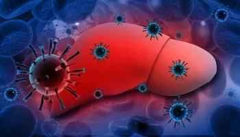
Interrupting nucleos(t)ide analogue therapy in hepatitis B e antigen-negative disease is one strategy that achieves a functional cure, meaning loss of hepatitis B surface antigen (HBsAg), in up to 20% of patients. It also carries a risk of viral rebound and alanine aminotransferase flares severe enough to be life-threatening, which makes picking the right candidates the whole problem.
How often that pays off at scale comes from RETRACT-B, an international cohort of 1552 virally suppressed patients who stopped therapy.2 Cumulative HBsAg loss reached 3.2% at 12 months and 13.0% at 48 months, short of the 20% ceiling drawn from selected cohorts. The strongest reported predictor was quantitative HBsAg at the end of treatment, with levels below 100 IU/mL carrying a subdistribution hazard ratio (sHR) of 22.5 (95% CI, 13.1-38.7) against higher levels. Loss also ran higher in White than Asian patients (sHR, 6.8; 95% CI, 2.7-16.8), with the authors recommending a stop threshold of under 1000 IU/mL for White patients and under 100 IU/mL for Asian patients. Hepatic decompensation ran 0.48 per 1000 person-years.
Those viral markers describe the virus rather than the response to it. Hepatitis B core-related antigen and HBV RNA have been proposed as additions, and earlier work in this cohort found that HBV-specific T-cell responses tracked sustained viral control but failed to distinguish loss of surface antigen from persistence.1 That gap sent the investigators to the B cells.
Participants had hepatitis B e antigen-negative disease with undetectable HBV DNA and normal alanine aminotransferase for at least 3 years on therapy; advanced fibrosis, cirrhosis, hepatocellular carcinoma, immunosuppression, and viral coinfection were exclusions. The cohort was 81% male, with a median age of 57 years and a median of 8 years of treatment, 76% of it tenofovir and 24% entecavir. Each underwent liver biopsy at the end of treatment, with blood sampled at weeks 12 and 48 and, in 7 patients, 4 to 8 years out.
Outcomes split 3 ways. Five patients (24%) lost HBsAg and seroconverted to anti-HBs; 11 kept the virus controlled without restarting therapy; and 5 required re-treatment after both an alanine aminotransferase flare and DNA rebound.
Prolonged therapy had already flattened a difference seen in untreated disease, where core-specific memory B cells outnumber surface-specific ones 5- to 18-fold. In this cohort the 2 populations were present at comparable frequencies with equivalent class switching, though the surface-specific cells still carried more programmed cell death protein 1 (PD-1), the surface marker that tracks with the functionally defective phenotype seen in chronic infection.
At the end of treatment, activated and class-switched cells within the global memory B-cell pool were significantly, if subtly, enriched in the patients who would go on to clear HBsAg. Frequencies of the antigen-specific populations themselves showed no such difference, which matters because the global measurement is the one a standard flow cytometry panel could deliver.
By week 48 the separation had widened. PD-1 on atypical surface-specific memory B cells fell in everyone at week 12, then rebounded where the antigen persisted while staying negligible in those who cleared it. Activation markers and plasmablasts rose selectively in the clearance group, circulating T follicular helper cells correlated tightly with surface-specific memory B cells there, and functional testing showed those cells could differentiate into antibody-producing cells much as cells from vaccinated controls did. Patients with no reduction in quantitative HBsAg produced negligible or undetectable responses. Anti-HBs levels between 20 and 1000 IU/mL and the B-cell changes were still present 4 to 8 years later.
Core-specific memory B cells behaved as a separate signal, rising a mean fourfold between the end of treatment and week 12, from under 1% to above 4% of the memory pool in 4 patients and to some degree in 12 of the remaining 17. Frequencies came back down by week 48 for most patients regardless of outcome.
The link to flares was real but loose. Increases were more common in patients with alanine aminotransferase above twice the upper limit of normal, yet some rose without any detectable flare and others held flat through a marked one. That inconsistency, and the fact that only 5 patients cleared HBsAg, is why the authors labeled the work exploratory and called for larger cohorts before any of it informs a stopping decision.
The practical appeal is straightforward even so. A marker read at the end of treatment, before a patient absorbs the risk of withdrawal, beats one read afterward, and the activated memory B-cell fraction is the kind of measurement a hospital flow cytometry laboratory already performs. Whether it beats quantitative HBsAg is for a larger trial to settle.
The study received antigen reagents from Gilead Sciences, which employs 2 of the study's authors; other authors disclose ties to Gilead and AbbVie.
References
1. Lens S, Burton AR, Davies J, et al. Tracking B cell immunity during perturbation of hepatitis B infection induced by treatment withdrawal. Gut. 2026;75(8):1587-1596. doi:10.1136/gutjnl-2024-333309
2. Hirode G, Choi HSJ, Chen CH, et al. Off-therapy response after nucleos(t)ide analogue withdrawal in patients with chronic hepatitis B: an international, multicenter, multiethnic cohort (RETRACT-B study). Gastroenterology. 2022;162(3):757-771.e4. doi:10.1053/j.gastro.2021.11.002